Pancreas anatomy
Click to enlarge
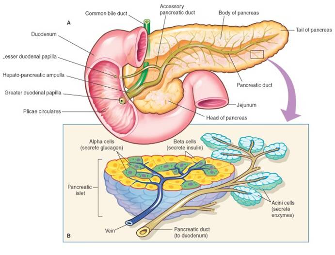
The pancreas is a retroperitoneal structure which extends transversely across the posterior abdominal wall from the duodenum to the spleen (see figure). It consists of both exocrine and endocrine tissues. The endocrine cells of the pancreas are found in the islets of Langerhans which are compact spherical masses embedded within the acinar tissue (exocrine). Each of the 1 million islets found in the pancreas measures between 100-200µm in diameter and consists of hundreds of endocrine cells. There are several types of endocrine cells: α-cells which secret glucagon, β-cells secreting insulin, δ-cells secreting somatostatin, and enterochromaffin cells which secret pancreatic polypeptide and digestive polypeptide hormones respectively.
β-cell and insulin

Click to enlarge
β cells make up of 65-80% of the islet cells and are solely responsible for the production and secretion of insulin, a hormone responsible for the regulation of plasma glucose in the body. Being a peptide, insulin is formed through gene transcription, translation and post-translational modification. mRNA is first translated into the inactive preproinsulin, whose signal sequence at the N-terminus allows it to then migrate out of the nucleus and into the endoplasmic reticulum (ER). Once arrived in the ER, the signal sequence is removed from preproinsulin to form the still inactive proinsulin. Through further di-sulfide bond formation between chain A and chain B, as well as the cleavage of chain C, pro-insulin is now transformed into active insulin. Insulin is stored in secretory granules in β-cells.
As blood glucose level increases, glucose from nearby blood vessels enter β cells via the type 2 glucose transporter GLUT2. Undergoing glycolysis in the cytoplasm and later respiration in the mitochondria, glucose increase ultimately results in ATP production and thus the increase of ATP/ADP ratio. This increase in ATP/ADP ratio induces the closure of ATP-sensitive potassium channel, preventing the outward migration of potassium ions. As a result, a positive charge rises and causes depolarisation. The voltage-gated calcium channels nearby respond to depolarisation by transporting calcium ions into the cell. The increase in intracellular calcium concentration results in the exocytosis of insulin from the secretory granules into the outside of β cells. Insulin is then assisted in its diffusion into the nearby blood vessels and travel to different places in the body (especially skeletal muscles and adipose tissues) to initiate plasma glucose uptake.
As blood glucose level increases, glucose from nearby blood vessels enter β cells via the type 2 glucose transporter GLUT2. Undergoing glycolysis in the cytoplasm and later respiration in the mitochondria, glucose increase ultimately results in ATP production and thus the increase of ATP/ADP ratio. This increase in ATP/ADP ratio induces the closure of ATP-sensitive potassium channel, preventing the outward migration of potassium ions. As a result, a positive charge rises and causes depolarisation. The voltage-gated calcium channels nearby respond to depolarisation by transporting calcium ions into the cell. The increase in intracellular calcium concentration results in the exocytosis of insulin from the secretory granules into the outside of β cells. Insulin is then assisted in its diffusion into the nearby blood vessels and travel to different places in the body (especially skeletal muscles and adipose tissues) to initiate plasma glucose uptake.
Insulin signalling and action

Click to enlarge
At target cells and tissues, insulin binds to tyrosine kinase insulin receptors, whose intracellular tyrosine kinase domain begins signal transduction by phosphorylating the scaffold protein IRS-1 which in turn activates PI3 kinase. This activation initiates the Akt signalling pathway by bringing forth the binding of Akt to PDK1 and PDK2. Akt is activated through the conformational change brought by the negative charge in its binding with the PDK’s. Once activated, Akt phosphorylates AS160, a Rab GTPase-activating protein, responsible for the trafficking process of vesicles containing glucose transporter GLUT4 from the ER to the cell membrane. The deployment of GLUT4 allows glucose to be taken into the cells in skeletal muscles and adipose tissues, preventing the high glucose plasma level.
T.N. Budi A.N. Khuong C. April | Copyright ©2012
